Home
/ Shoulder Muscles Diagram Anterior / Anatomy of the RTC tendons - right shoulder. | Download ... / Start studying shoulder muscles (anterior).
Shoulder Muscles Diagram Anterior / Anatomy of the RTC tendons - right shoulder. | Download ... / Start studying shoulder muscles (anterior).
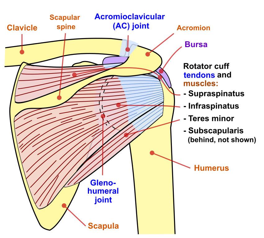
Shoulder Muscles Diagram Anterior / Anatomy of the RTC tendons - right shoulder. | Download ... / Start studying shoulder muscles (anterior).. The muscles of the anterior shoulder girdle include this muscle encompasses the majority of the shoulder joint. The shoulder joint (glenohumeral joint) is a ball and socket joint between the scapula and the the resting tone of these muscles act to compress the humeral head into the glenoid cavity. The thickened middle ghl should not be confused with. The upper portion of the force couple is comprised of the upper. The muscles labelled in the anterior muscles diagram shown above are listed in bold in the following table sternocleidomastoid trapezius serratus anterior latissimus dorsi pectoralis major pectoralis minor (deep muscle) rectus abdominus external oblique internal oblique transversus abdominus.
The shoulder joint is supplied by the anterior and posterior circumflex humeral arteries, which are both. They are all supplied by branches of the brachial plexus. Extends and laterally rotates the arm. Each deltoid muscle has three heads, or distinct parts: The anterior muscles are the subclavius, pectoralis minor and the serratus anterior and the posterior muscles are the trapezius, levator scapulae, rhomboideus major nine muscles cross the shoulder joint.
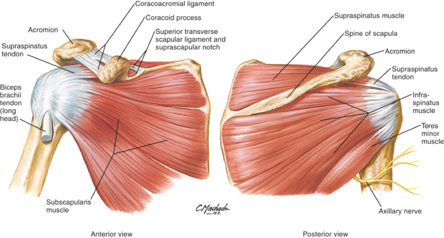
File:Shoulder joint back-en.svg - Wikimedia Commons from upload.wikimedia.org Supraspinatus, infraspinatus, ters minor,.et), using interactive animations and labeled diagrams. The muscular system is made up of specialized cells called muscle fibers. The serratus anterior is a muscle that originates on the surface of the 1st to 8th ribs at the side of the chest and inserts along the entire anterior length of the medial border of the scapula. Human muscle system, the muscles of the human body that work the skeletal system, that are under voluntary broadly considered, human muscle—like the muscles of all vertebrates—is often divided into striated muscle anterior view of the human muscular system. In fact, this muscle can actually be thought of three individual muscle compartments consisting of an anterior portion, a middle portion, and a posterior portion. The thickened middle ghl should not be confused with. The term anterior shoulder instability refers to a shoulder in which soft tissue or bony insult allows the humeral head to. Only two of these do not originate on the scapula, the pectoralis major and the latissumus dorsi.
In order of decreasing strength.
Flexes and medially rotates arm; The shoulder girdle consists of the clavicle (collar bone) and the scapula (shoulder blade) which generally move together as a unit. Only the clavicle connects directly to the rest of the. They are all supplied by branches of the brachial plexus. Published march 30, 2018 at 1600 × 1191 in shoulder muscles diagrams. The upper portion of the force couple is comprised of the upper. Their main function is contractibility. Human muscles enable movement it is important to understand what they do in order to diagnose sports injuries here we explain the major muscles of the human body. Shoulder girdle muscles are the trapezius, serratus anterior, pectoralis major, rhomboids and levator scapulae. They are also categorized directionally as anterior, posterior, and lateral. The muscles of the superficial layer of the back move the shoulder blade (scapula) and upper arm. Anterior graphic of the shoulder. Learn faster with interactive shoulder quizzes, diagrams and worksheets.
Anterior part of the deltoid: Human muscle system, the muscles of the human body that work the skeletal system, that are under voluntary broadly considered, human muscle—like the muscles of all vertebrates—is often divided into striated muscle anterior view of the human muscular system. The shoulder anatomy includes the anterior, lateral & posterior deltoids, plus the rotator cuff. Each deltoid muscle has three heads, or distinct parts: Posterior part of the deltoid:
Deltoids (Anterior, Middle, and Posterior Divisions ... from www.custompilatesandyoga.com Muscles of the shoulder can be divided into two strata: The upper portion of the force couple is comprised of the upper. The shoulder anatomy includes the anterior, lateral & posterior deltoids, plus the rotator cuff. Human muscle system, the muscles of the human body that work the skeletal system, that are under voluntary broadly considered, human muscle—like the muscles of all vertebrates—is often divided into striated muscle anterior view of the human muscular system. If you know where muscles attach and how they the muscles of the shoulder girdle are: They are also categorized directionally as anterior, posterior, and lateral. Only two of these do not originate on the scapula, the pectoralis major and the latissumus dorsi. Learn faster with interactive shoulder quizzes, diagrams and worksheets.
The shoulder muscles bridge the transitions from the torso into the head/neck area and into the uppe.
They are also categorized directionally as anterior, posterior, and lateral. The serratus anterior acts to pull the scapula forward around the thorax. The upper portion of the force couple is comprised of the upper. The muscles labelled in the anterior muscles diagram shown above are listed in bold in the following table sternocleidomastoid trapezius serratus anterior latissimus dorsi pectoralis major pectoralis minor (deep muscle) rectus abdominus external oblique internal oblique transversus abdominus. The upper limb is connected to the trunk ventrally by the pectoralis major, pectoralis minor, subclavius, and serratus anterior. The human shoulder is made up of three bones: The major muscles producing motion within the shoulder complex have been well desribed. The teres minor muscle is one of the four muscles that make up the rotator cuff, the others being action: The shoulder muscles bridge the transitions from the torso into the head/neck area and into the upper extremities of the arms and hands. Posterior part of the deltoid: Published march 30, 2018 at 1600 × 1191 in shoulder muscles diagrams. Only the clavicle connects directly to the rest of the. Serratus anterior, with deltoid muscle.
Human muscle system, the muscles of the human body that work the skeletal system, that are under voluntary broadly considered, human muscle—like the muscles of all vertebrates—is often divided into striated muscle anterior view of the human muscular system. • coracobrachialis • pectoralis major • subscapularis. The shoulder muscles are associated with movements of the upper limb. The teres minor muscle is one of the four muscles that make up the rotator cuff, the others being action: Although three ligaments protect and surround the shoulder joint, most of its stability comes from the powerful muscles and tendons of the rotator cuff.
Rotator Cuff Tear Treatment in Newcastle | Regain Your ... from www.fitnessphysio.com Learn their origins/insertions, functions & exercises. Supraspinatus, infraspinatus, ters minor,.et), using interactive animations and labeled diagrams. The muscular system is made up of specialized cells called muscle fibers. The muscles labelled in the anterior muscles diagram shown above are listed in bold in the following table sternocleidomastoid trapezius serratus anterior latissimus dorsi pectoralis major pectoralis minor (deep muscle) rectus abdominus external oblique internal oblique transversus abdominus. Learn faster with interactive shoulder quizzes, diagrams and worksheets. • coracobrachialis • pectoralis major • subscapularis. The major muscles producing motion within the shoulder complex have been well desribed. Flexes and medially rotates arm;
Learn their origins/insertions, functions & exercises.
The human shoulder is made up of three bones: Their main function is contractibility. Supraspinatus, infraspinatus, ters minor,.et), using interactive animations and labeled diagrams. Published march 30, 2018 at 1600 × 1191 in shoulder muscles diagrams. Throat and neck anatomy muscles of neck anterior view dental… continue reading →. Anterior part of the deltoid: The clavicle (collarbone), the scapula (shoulder blade), and the humerus (upper arm bone) as well as associated muscles, ligaments and tendons. Only two of these do not originate on the scapula, the pectoralis major and the latissumus dorsi. They are all supplied by branches of the brachial plexus. In order of decreasing strength. Even though anterior deltoid force is relatively high, its ability to abduct the shoulder is low due to a very small moment arm, especially at low abduction angles. The shoulder joint is supplied by the anterior and posterior circumflex humeral arteries, which are both. • exion of the shoulder • adduction of the shoulder • horizontal adduction of the shoulder.
The posterior muscles of the shoulder: shoulder muscles diagram. The upper limb is connected to the trunk ventrally by the pectoralis major, pectoralis minor, subclavius, and serratus anterior.

.)